Endodontic Treatment ( Root Canal Therapy )
Tooth Anatomy
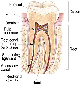
Endodontics is the Specialty of
Dentistry that involves the treatment and prevention of
pulpal and periradicular (structures surrounding the root)
disease. Endodontic treatment or
tooth extraction is required when the tooth's pulp tissue
has developed irreversible disease. |
What causes pulpal disease?
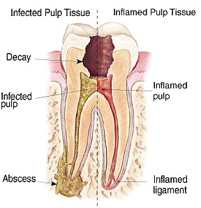 Endodontic disease is strictly a microbial disease. It results from bacteria, yeast and viral infection of the pulp tissue and spaces. The most common pathway for the microbial invasion is past and/or current cavities. Cracks in the crown and/or the root can also let microbes invade the pulp spaces. Traumatic blows to the tooth can sometimes damage the blood supply of the pulp, resulting in death of the pulp. In some rare instances of advanced gum disease, the loss of support and seal that is normally provided by the periodontal ligament can result in pulpal infection. |
How is Endodontic Treatment Performed?
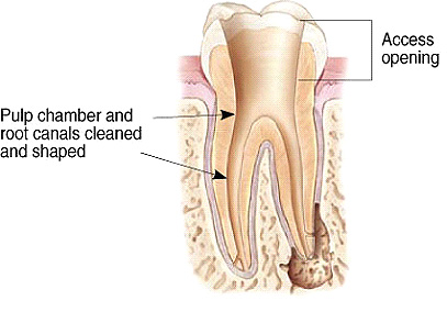 |
After profound anesthesia is obtained,
the
tooth is isolated from the oral cavity and saliva by
covering it with a rubber sheet (rubber dam). The decay is removed, the pulp spaces are located and mechanically sculpted to remove the diseased pulp tissue, and to facilitate the flow and placement of disinfectants. The instrumentation phase is the most technically challenging aspect of treatment. The canals are usually quite narrow, and have marked curvatures, with frequent spiral and branching configurations. Commonly, the openings to some of the canals have closed and careful exploration is required to access and negotiate the canal spaces. |
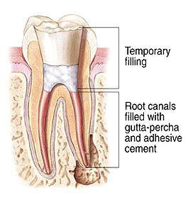 |
After disinfection of the tooth is completed the root canal spaces are filled with a natural rubber material (Gutta Percha) and a cement sealer. We generally place a temporary filling at the end of our treatment, and your restorative dentist restores the crown of the tooth. It is critical that you promptly return to you restorative dentist to have the crown of the tooth restored. The outcome of Endodontic treatment depends equally upon the quality of the Restorative and Endodontic treatment. All temporary filling materials and root canal filling materials will, over time, allow microbial penetration. The prevention of re-infection of the canal spaces depends upon timely and adequate restoration of the crown of the tooth. |